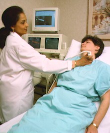
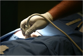
Welcome to SonoNet's patient resource page.
SonoNet is committed to providing the highest quality in care to our patients.
We continually refine the care we provide, we monitor and measure the treatments
our patients receive and evaluate our performance against our own rigorous
standards as well as industry benchmarks.
At SonoNet, we define quality as:
- Superior Care and Outcomes
- Outstanding Patient Care
- Excellent Service and Patient Satisfaction
- Physician & Patient Education
Information
for Patients
What
is Ultrasound
We're
leaders in treating as well as preventing — the most complex cardiovascular
conditions. . We strive to better serve patients, to improve the health care
experience.
What
Is Ultrasound
Ultrasound
(US) imaging, also called ultrasound scanning or sonography, is a method of
obtaining images from inside the human body through the use of high frequency
sound waves. The reflected sound wave echoes are recorded and displayed as
a real-time visual image. Ionizing radiation (x-rays) are not involved in
ultrasound imaging.
Ultrasound
is a useful examination tool for many of the body's internal organs, including
the heart, liver, gallbladder, spleen, pancreas, kidneys, and bladder. Because
ultrasound images are captured in real-time, they can show movement of internal
tissues and organs, and enable physicians to see blood flow and heart valve
functions.
What
is Diastolic Dysfunction
Aging
and most diseases of the heart affect the ability of the heart to relax and
fill properly because the muscles become stiffer. In most of these cases the
basic units of the heart, the muscle cells, die and are replaced by cells
that are not elastic. This loss of elasticity causes stiffening of the heart
and is a progressive process.
What
Is Diastolic Dysfunction
Information
About Exams
For
specific information about an exam we perform Select
Exam to view more detailed info about exam.
Echocardiogram
Facts - Questions & Answers
Q:
What is an echocardiogram?
A: An echocardiogram is a safe, non-invasive procedure used to diagnose cardiovascular
disease. By using echocardiography to visualize anatomy, structure, and function,
doctors can quickly diagnose the presence and severity of heart valve problems,
or determine abnormal flow within the heart which occurs with congenital heart
disease. An echocardiogram provides your doctor with a non-invasive window
to your heart, and enables a cardiologist along with your Doctor to diagnose
a number of cardiovascular diseases and prescribe proper treatment.
Q. How does it work?
A: Echocardiograms reflect high-frequency sound waves directly off the heart
tissue to create images of its structure: its four chambers, heart valves,
the great blood vessels entering and leaving the heart, as well as the sac
around the heart.
SonoNet
has a team of medical imaging specialists who devote themselves to bringing
the best evidence based medicine to patients. Their goal is to help separate
wellness from disease and provide the best cardiovascular care.
Our
Team of Physicians -


