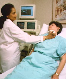
Vascular ultrasound is a noninvasive
ultrasound method used to examine the blood circulation in the arms and
legs. Non- invasive means the procedure does not require the use of needles,
dyes, radiation or anesthesia.
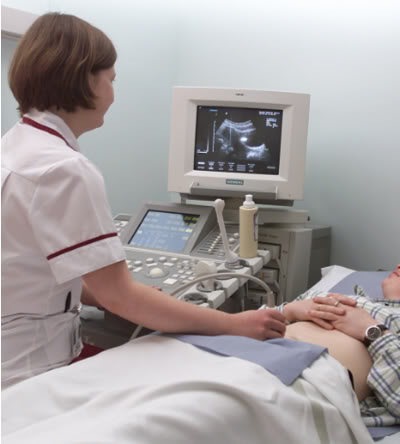
During a vascular ultrasound, sound
waves are transmitted through the tissues of the area being examined.
These sound waves reflect off blood cells moving within the blood vessels,
allowing the reading physician to calculate their speed.
Carotid
Duplex Ultrasound:
Carotid Ultrasound
with Doppler – This exam
uses reflected sound waves to visualize the right and left common carotid
arteries from the base of the neck to just above the bifurcation of the
internal and external carotid arteries. The vertebral artery (posterior
in the neck) is also imaged. The physician evaluates the images to determine
the extent of any blockage to these arteries. Doppler is used to show how
much blood is flowing to the brain and eyes. The
length of this test is 45 minutes.
No preparation is needed.
*
Symptoms: cervical or carotid bruit, memory loss, cluster
type headache, vertigo, aphasia/dysphasia, previous stroke, motor or sensory
deficit, syncope, fluctuating confusion, Amaurosis Fugax (transient monocular
blindness), unilateral paralysis/weakness, drop attacks, and coronary
or peripheral artery disease.
Lower
Extremity Arterial Ultrasound:
Lower Extremity
Arterial Imaging – This
exam evaluates the arterial blood flow from the pelvis to the foot via reflected
sound waves. A Cardiologist analyzes the images and Doppler waveforms to
determine the location and extent of blockages. This
exam takes approximately 45 minutes per leg.
No preparation is needed. For best comparative results, both legs should
be scanned.
*
Arterial Symptoms: claudication, leg pain, rest pain, bruits,
gangrene, diabetic neuropathy, skin color changes or ulceration, absent
or diminished distal or pedal pulses, distal extremity hair loss, skin
or nail infections, hypertension, and extreme weakness or fatigue.
Lower
Extremity Venous Ultrasound:
Lower Extremity Venous
Imaging – This exam uses
sound waves to visualize the veins from the pelvis to the foot. Doppler
is used to evaluate blood flow in the veins. The physician views these images
to determine the presence of a blood clot or venous abnormality. This
exam takes approximately 45 minutes per leg.
There is no preparation for this exam. Please specify which leg
or both.
*
Venous Symptoms: edema, pitting edema, pain, increased
limb tenderness, anti-coagulant therapy evaluation, skin discoloration,
ulcers, varicose veins and pulmonary embolism.
Upper
Extremity Venous or Arterial Ultrasound:
Upper
Extremity Venous or Arterial Imaging –
These exams use reflected sound waves and Doppler to evaluate the veins
or arteries in the arm. A physician indicates which tests are needed. The
Upper Extremity Venous visualizes the presence of a blood clot. The Upper
Extremity Arterial can determine the severity of an arterial blockage. This
testing takes less than one hour. No preparation is needed. (Physician
specifes which arm, or both.)
*
Venous Symptoms: Edema, pain- tenderness, ulcers
*
Arterial Symptoms: arm pain, skin or nail infections,
skin color changes or ulceration, absent or diminished pulses, gangrene,
numbness and positive Allen's test.
Abdominal
Aortic Aneurysm (AAA) Ultrasound:
Abdominal
Aortic Aneurysm (AAA) –
This exam uses ultrasound to evaluate the Abdominal Aorta or signs of a
potential Aortic Aneurysm. Aortic aneurysms can develop anywhere along the
length of the aorta. The majority, however, are located along the abdominal
aorta. Most (about 90%) of abdominal aneurysms are located below the level
of the renal arteries, the vessels that leave the aorta to go to the kidneys.
About two-thirds of abdominal aneurysms are not limited to just the aorta
but extend from the aorta into one or both of the iliac arteries.
*
Symptoms: Most abdominal aortic aneurysms produce no
symptoms (they are asymptomatic).
Abdominal
aortic aneurysm can remain asymptomatic or produce mild to moderate symptoms
for years. However, a rapidly expanding abdominal aneurysm can cause sudden
onset of severe, steady, and worsening middle abdominal and back pain